A broken jaw does not automatically require a plate. The right choice depends on the fracture’s location and stability, your pre-injury bite, CT findings, nerve and eye function, infection risk and ability to complete follow-up. By the end, you will know when observation, jaw immobilisation, miniplates or a stronger reconstruction plate is usually considered, and what questions to ask before treatment.
Key takeaways
- Mandible fractures may need stable fixation to restore bite, chewing and facial symmetry.
- Small, stable fractures can sometimes heal without plates when the bite remains aligned.
- Choose fixation based on fracture location, displacement, bone quality and chewing forces.
- Follow-up checks bite, infection, mouth opening, bone healing and whether plate removal is needed.
Which facial bones are involved, and what must reconstruction restore?
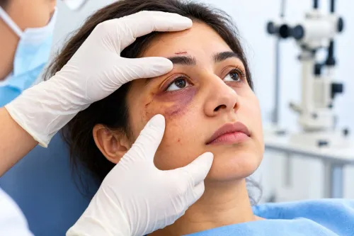
Jaw fracture reconstruction in Chennai begins by identifying whether the injury affects the mandible, maxilla, cheekbone, orbital rim or several facial bones together. Mandibular fractures may involve the tooth-bearing body, angle, symphysis, ramus or condyle.
| Bone or region | What can be disrupted | What reconstruction must protect |
|---|---|---|
| Mandible | Bite, ramus height, chewing and lower-lip sensation | Pre-injury occlusion and jaw movement |
| Maxilla or Le Fort fracture | Upper dental arch and midface buttresses | Facial height, projection and bite |
| Zygomatic bone | Cheek width and orbital boundary | Facial symmetry and eye position |
| Orbital rim or floor | Orbital volume and eye movement | Globe position and single vision |
| Multiple facial bones | Several facial planes at once | Coordinated alignment across the face |
Zygomatic and orbital-rim fractures can alter facial width, eye position or orbital volume even when your teeth meet normally. Facial fracture surgery in Chennai therefore requires more than checking the mandible.
A CT scan of the facial bones, often with three-dimensional reconstructions, shows displacement, comminution and orbital involvement more clearly than plain X-rays. Match it with examinations of your bite, eye movements, facial sensation and facial movement, and record pre-injury occlusion from dental records, photographs or family information when available.
Once you are medically stable, assess the bite before swelling settles and repeat it during follow-up. The goals are to restore your pre-injury bite, facial height, symmetry and function—not simply to insert metal. A new open bite, crossbite or shifted midline signals that reduction or fixation needs review.
When is a plate necessary, and when can treatment avoid one?
A plate is not necessary for every jaw fracture. A nondisplaced or favourably aligned fracture can heal with observation, a soft diet and scheduled examinations when the bite remains unchanged and the fracture is stable.
Closed treatment uses intermaxillary fixation—wires, screws, elastics or dental bars—to hold the upper and lower jaws together temporarily. It suits selected fractures when restoring the bite aligns the fragments and you can manage restricted eating, oral hygiene and follow-up.
Open reduction and internal fixation is more likely for a displaced, unstable, open, infected or multiple fracture, or one where a small shift would change chewing or facial shape. A break communicating with the mouth, including many fractures through tooth-bearing mandibular bone, is treated as contaminated or open in clinical decision-making.
Dental infection, a devitalised tooth, smoking and poor oral hygiene increase the risks of infection, nonunion and plate exposure. Antibiotics do not replace reduction, debridement when indicated, dental care and stable fixation.
Condylar and subcondylar fractures are not automatically plated. Occlusion, ramus height, displacement, mouth opening, associated fractures and your ability to rehabilitate determine whether functional treatment, temporary immobilisation or open fixation is safer.
For maxillofacial trauma reconstruction in Chennai, ask a jaw fracture surgeon in Chennai:
- How will bite stability and fracture movement be checked?
- Is the fracture open or contaminated through the mouth?
- What eating, hygiene and rehabilitation restrictions apply?
- What finding would make fixation or plate removal necessary?
Which fixation option matches the fracture’s mechanical demands?
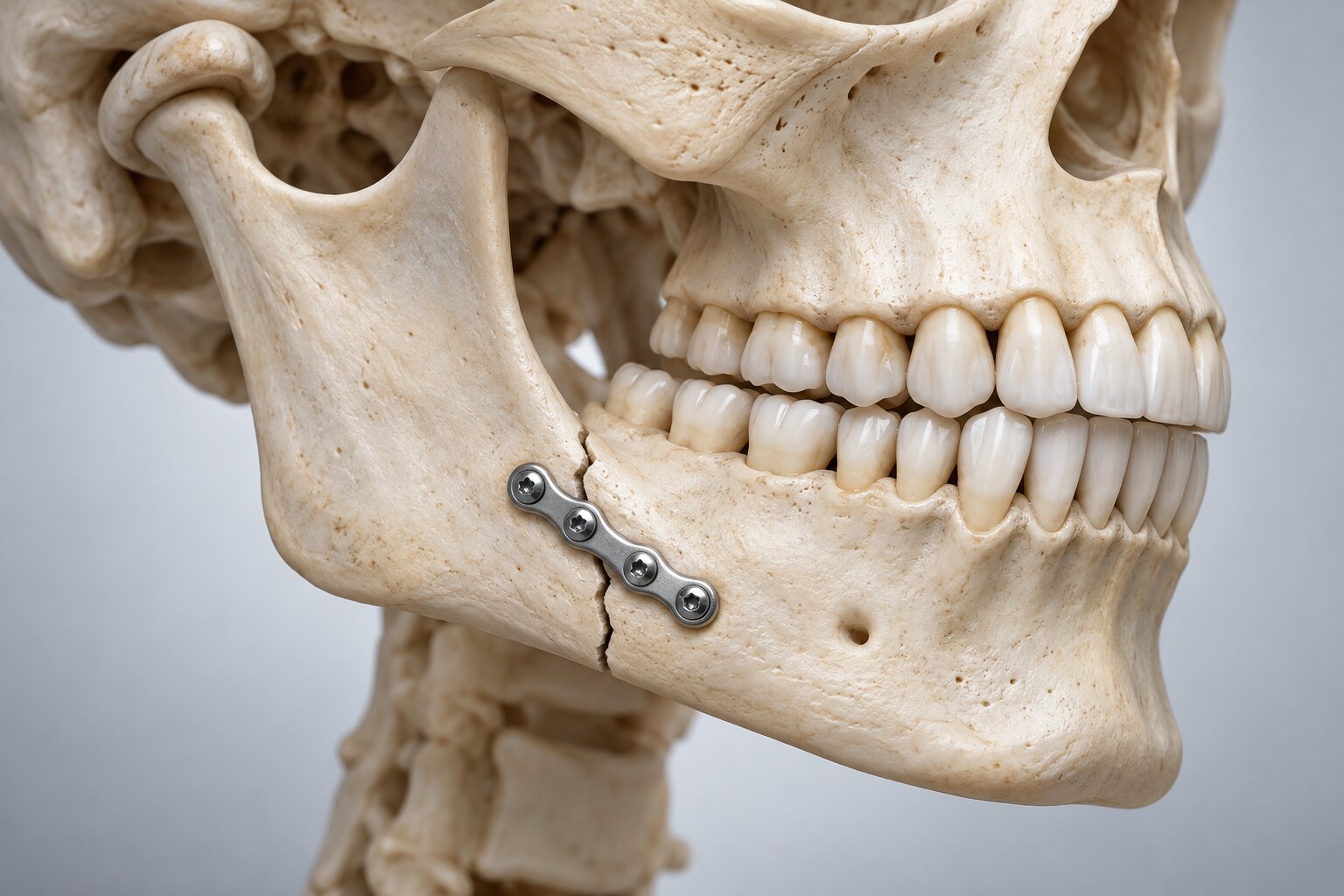
Choose fixation by asking whether the bone can share chewing force. Titanium miniplates and screws suit well-aligned fragments with broad bony contact; reconstruction plates act as load-bearing bridges when that contact cannot be trusted.
| Option | Mechanical role | When it fits | Main limitation |
|---|---|---|---|
| Titanium miniplate and screws | Load-sharing | Stable, well-aligned fractures | Unsuitable for large gaps or severe comminution |
| Reconstruction plate | Load-bearing bridge | Bone loss, comminution, severe mandibular atrophy or poor contact | Wider exposure and more soft-tissue irritation |
| Resorbable plate | Temporary support without permanent metal | Selected fractures with lower loading demands | Limited strength, degradation and handling range |
| Intermaxillary fixation | Holds the jaws together | Closed treatment or added support | Restricts speech, eating, hygiene and airway access |
| Observation and soft diet | Allows stable bone to heal without fixation | Nondisplaced fractures with an unchanged bite | Requires close review; movement or bite change can alter the plan |
More metal is not automatically safer. An unnecessarily large plate can require wider exposure, irritate tissue and complicate later treatment. Screw length and plate position must protect tooth roots, the inferior alveolar nerve and the fracture line.
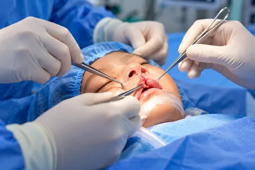
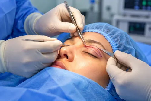
Intraoral access avoids an external scar but places the incision in a bacteria-rich environment and limits access for some injuries. External access improves visibility in complex facial fracture surgery in Chennai, but adds a scar and route-specific risks, including facial-nerve injury. These trade-offs matter when planning jaw fracture reconstruction in Chennai.
How are complex midface and accident injuries reconstructed?
1. Facial reconstruction after accident in Chennai may require staged treatment, not one operation. Emergency care addresses the airway, major bleeding, brain injury and eye-threatening damage. Definitive bone reconstruction follows medical stabilisation and a complete injury assessment, often using CT to map displacement, comminution and orbital involvement.
2. In Le Fort and other maxillary fractures, surgeons establish dental occlusion—the relationship between the upper and lower teeth—before rebuilding the facial buttresses supporting the nose, cheek and midface. Ignore that sequence and fixation can leave a persistent open bite or abnormal facial projection.
3. Complex maxillofacial trauma reconstruction in Chennai may involve the mandible, maxilla, zygomatic bones, orbital rims and nasal structures together. A stable mandible alone cannot correct double vision, a sunken eye, midface flattening or a displaced cheekbone.
Orbital repair therefore assesses eye movement, globe position and orbital volume, while coordinated fixation prevents facial segments from healing in different planes.
4. Record lower-lip and chin sensation before surgery because inferior alveolar or mental-nerve injury from the accident may persist after technically successful fixation. Choose an intraoral or external approach by balancing direct visibility against scar exposure, oral contamination and nerve risk.
What follow-up determines healing, mouth opening and plate removal?
Healing is checked through your bite, pain, infection signs, fragment mobility and imaging when indicated. A new open bite, crossbite or shifted midline needs prompt review; a plate cannot correct movement by itself.
Once the surgeon confirms that the fracture is stable, begin graded jaw-opening and functional exercises, especially after a condylar injury. Do not force the jaw early or resume hard chewing without clearance: excessive load can loosen fixation or disturb healing.
Report these changes promptly:
- Fever, increasing swelling, pus or a foul taste
- Worsening pain or new mobility at the fracture site
- A plate visible through the mouth
- A new bite change
- Persistent or worsening numbness of the lower lip or chin
Titanium plates often remain permanently when covered, painless and unrelated to symptoms. Routine removal is not universally required. Surgeons consider removal for infection, exposure, persistent pain, loosening, prominent hardware or interference with function. Removal is another operation with anaesthesia, bleeding, nerve and infection risks, so its benefit must outweigh those risks.
When comparing a jaw fracture surgeon in Chennai, ask how the plan uses your pre-injury bite, CT findings, nerve status, oral contamination, plate load requirements, rehabilitation and removal criteria. If you are assessing facial reconstruction after accident in Chennai, The Face Clinic can help structure those questions around the documented injury and proposed treatment plan.
Frequently asked questions
Which facial bones can be involved in a jaw fracture?
Injuries may affect the mandible, maxilla, cheekbone, orbital rim or multiple facial bones. Mandibular fractures include the body, angle, symphysis, ramus and condyle.
When can a jaw fracture heal without a plate?
Non-operative treatment can be considered when the fracture is stable, minimally displaced and the bite remains aligned. The decision also depends on pain, mouth opening, infection risk and follow-up findings.
How do surgeons choose between plates, screws and other fixation?
Fixation matches the fracture’s location, displacement, bone quality and mechanical load. Options include miniplates, stronger load-bearing plates, lag screws, external fixation and conservative immobilisation.
What does follow-up after facial fracture surgery check?
Follow-up evaluates bite alignment, wound healing, infection, facial sensation, mouth opening, chewing function and bone union. Imaging helps identify delayed healing or hardware problems.
Keywords