Eye-area surgery is not a single procedure: the pathway differs for a fractured orbit, a drooping or damaged eyelid, a blocked tear duct and an orbital mass. You will learn how assessment, scans, treatment decisions, anaesthesia, surgery, discharge and follow-up fit together, and which symptoms require urgent care.
Key takeaways
- Your first assessment checks vision, eye movement, eyelids, tear drainage and nerve function.
- Scans help distinguish conditions needing surgery from those suitable for observation.
- Orbit and eyelid surgery steps depend on the affected tissue and surgical goal.
- Ask who coordinates your eye, facial nerve and reconstructive care after discharge.
Your First Consultation: Symptoms, Vision and Eye Movement
Understanding what happens during oculoplastic surgery in Chennai starts with an examination, not an operation. The surgeon first identifies whether your problem involves the eyelid, eye surface, tear drainage system, orbit, facial muscles or nerve function.
1. Describe your symptoms, including pain, watering, dryness, double vision, blurred vision, eyelid drooping, bulging, trauma and changes in appearance. State when each symptom began and whether it is worsening.
2. Bring a complete medical and medication history. Include aspirin, anticoagulants, antiplatelet medicines, supplements and diabetes treatment; do not stop any drug without instructions from its prescribing doctor and the surgical team.
3. Expect a vision check, pupil examination and eye-movement assessment. These checks can reveal restricted movement, double vision or pressure-related problems that an eyelid examination alone would miss.
4. The surgeon examines eyelid height, turning, closure, skin and muscle movement, facial sensation and asymmetry. They also check the tear film and ocular surface for dry eye, irritation or exposure from incomplete closure.
5. If watering is the complaint, fluorescein dye, punctum inspection, probing and irrigation help locate blockage in the punctum, canaliculus, lacrimal sac or nasolacrimal duct. Watery eyes can instead result from eyelid malposition or surface disease.
6. Clinical photographs and visual-field testing may document asymmetry or blocked peripheral vision. The findings determine whether the next step is observation, medication, an office treatment or further investigation—not surgery by default.
Scans and the Decision Between Observation and Surgery
A scan alone does not decide whether you need surgery. The oculoplastic surgery process in Chennai begins by matching symptoms and examination findings with the likely cause, then checking whether treatment can protect vision, relieve symptoms or correct appearance without an operation.
1. The surgeon reviews vision, pupil reactions, eye movements, eyelid position and closure, facial symmetry, tear-film condition and corneal exposure. Photographs and visual-field testing help document asymmetry, drooping or obstruction.
2. CT shows orbital bones, fracture lines, bleeding and bony expansion. MRI gives better detail of muscles, nerves, soft tissue and suspected orbital tumours. These scans are usually reserved for trauma, proptosis, thyroid eye disease, suspected tumour or unexplained findings, not routine cosmetic eyelid surgery.
3. The team chooses observation when the finding is stable, mild and not threatening vision. Medication may treat inflammation, thyroid eye disease, dry eye or infection; an office procedure may address a small lesion, injection need or selected tear-duct problem.
4. Surgery becomes appropriate when vision is threatened, eye closure is failing, pain or double vision persists, a tumour requires removal or tissue damage will not improve with simpler treatment. Tear testing, punctal examination, probing and irrigation distinguish duct blockage from eyelid malposition or ocular-surface disease.
The decision may involve an ophthalmologist, radiologist, anaesthetist, ENT specialist, pathologist or oncologist. Ask which finding changes the recommendation, what happens if you wait, and whether a second scan or biopsy is needed before scheduling an operation.
What Happens During Orbit and Eyelid Surgery
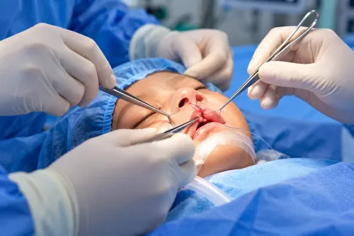
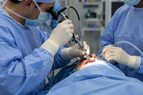
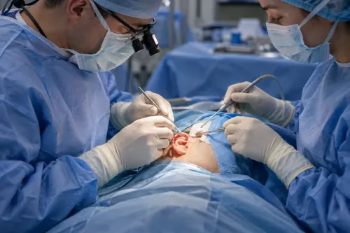
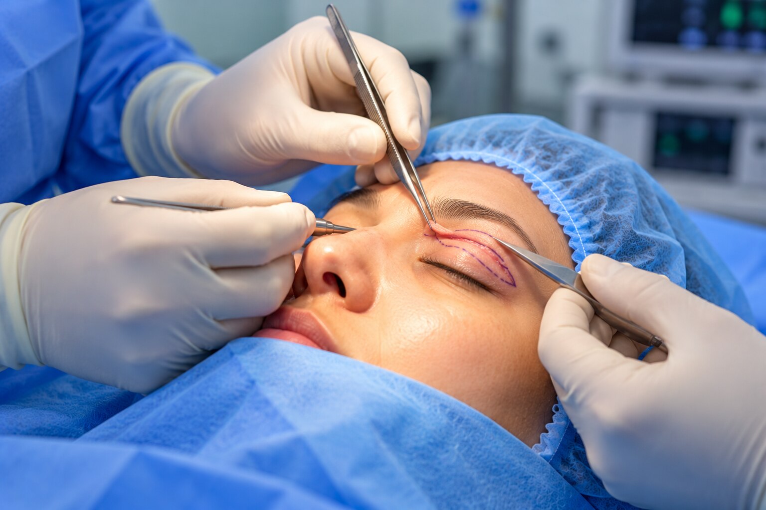
Orbit eyelid surgery steps begin with anaesthesia, skin marking and a final check of vision and eye movement. Local anaesthetic with intravenous sedation suits many eyelid operations; general anaesthesia is used for extensive orbital work, children or patients unable to tolerate local treatment.
- The surgeon chooses access through the eyelid crease, conjunctiva, outer orbital wall or nose, based on the structure involved.
- The abnormal tissue is repaired, removed or repositioned while protecting the eye, optic nerve and eye muscles.
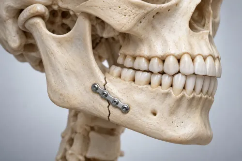
- Tumour specimens go to pathology; fracture repairs may use plates, screws or an implant to restore the orbital floor or wall.
- The incision is closed with sutures, and the team checks vision, bleeding and eye position before discharge or overnight observation.
| Operation | Main surgical aim | What makes it different |
|---|---|---|
| Fracture repair | Restore orbital volume and free a trapped muscle | Bone fragments are repositioned or reconstructed with an implant; double vision and facial numbness require monitoring |
| Tumour surgery | Remove or biopsy abnormal tissue safely | The approach depends on tumour location, and pathology determines further cancer treatment |
| Eyelid reconstruction | Rebuild the lid after cancer, trauma or tissue loss | Skin, muscle, lining and sometimes a graft or flap must be restored so the eye closes and remains protected |
Orbit decompression for thyroid eye disease removes selected bone or fat to reduce pressure or prominence, but it can cause new double vision, bleeding, sinus problems or altered sensation. Extensive cases need coordinated ophthalmic, anaesthetic, radiology, ENT and pathology support.
How Tear-Duct and Different Eyelid Operations Are Performed
Tear-duct surgery starts by locating the blockage, not by assuming watery eyes prove one exists. The surgeon examines the puncta, applies fluorescein dye, then probes and irrigates the drainage pathway to identify obstruction in the punctum, canaliculus, lacrimal sac or nasolacrimal duct. Eyelid malposition and ocular-surface disease can cause the same symptom.
If the duct is blocked near the nose, dacryocystorhinostomy creates a new passage from the lacrimal sac into the nasal cavity. The surgeon may perform it through a skin incision beside the nose or endoscopically through the nose, sometimes leaving a temporary silicone tube to keep the channel open.
Tear duct surgery in Chennai follows these same anatomical steps; complex cases may need ENT support.
Functional eyelid operations depend on the problem:
- Drooping: Levator advancement or resection suits useful levator muscle function; a frontalis sling connects the lid to the forehead muscle when levator function is poor.
- Turning inward or outward: The surgeon tightens, repositions or releases eyelid tissues so lashes stop rubbing the cornea or the lid contacts the eye properly.
- Scarring: Scar bands are released and replaced with grafted or rearranged tissue when they distort eyelid closure.
- Tissue loss: Local flaps, skin grafts or composite reconstruction restore the lid’s protective layers.
Local anaesthetic with sedation suits many operations; extensive reconstruction, childhood surgery or orbital involvement may require general anaesthesia. The team checks closure and vision before discharge.
Discharge, Recovery and Choosing Coordinated Care in Chennai
Expect bruising, swelling, tightness and temporary eyelid asymmetry after surgery. Ointment can blur vision, so use prescribed drops or ointment, cold compresses during the early period, head elevation and clean wound care. Avoid rubbing, strenuous activity and contact lenses until your surgeon permits them; drive only when your vision is clear and safe.
Seek emergency assessment immediately for:
- Rapidly increasing pain or swelling
- Reduced vision, a firm or protruding eye, or restricted eye movement
- Nausea or severe headache with orbital swelling
- Heavy bleeding, spreading redness, pus or fever
Do not wait for a routine appointment if these symptoms develop. Ask before discharge whom to call after hours and which hospital emergency department should assess you.
Choose a Chennai team by checking the coordination available for your diagnosis:
| Need | Team to confirm | Why it matters |
|---|---|---|
| Orbital tumour or unexplained proptosis | Ophthalmic surgeon, radiologist, pathologist and oncologist | Imaging and tissue diagnosis guide treatment |
| Trauma or complex tear-duct disease | Ophthalmic surgeon with ENT and maxillofacial support | Bone, nasal and drainage anatomy may require combined care |
| High-risk postoperative vision monitoring | Ophthalmic and anaesthetic teams with emergency access | Vision-threatening bleeding needs rapid treatment |
The Face Clinic is worth assessing by asking who handles pathology, imaging review and postoperative emergencies rather than choosing on procedure name alone. Provide the team with every medicine, including aspirin, anticoagulants, supplements and diabetes treatment; never stop one without instructions from the prescribing doctor and surgeon.
Frequently asked questions
What happens during the first oculoplastic consultation?
The surgeon reviews your symptoms and checks vision, eye movement, eyelids, the eye surface, tear drainage, facial muscles and nerve function.
When are scans needed before oculoplastic surgery?
Scans are used when the surgeon needs to assess the orbit, bones, soft tissues, a mass, trauma or deeper causes of eye-area symptoms.
What are the main steps in orbit and eyelid surgery?
The team confirms the surgical plan, gives anaesthesia, reaches the affected tissue through the planned incision, treats the problem, closes the wound and monitors you during recovery.
How is tear-duct surgery performed?
The surgeon opens or bypasses the blocked drainage pathway, often placing a temporary tube when needed, then checks drainage and provides aftercare instructions.
Keywords