A lump near the ear, swelling under the jaw, unexplained numbness, a non-healing extraction socket or an abnormal dental X-ray can signal a problem that routine dental treatment cannot classify. By the end, you will know which symptoms need urgent assessment, how salivary-gland and jaw lesions are investigated, and what to bring when you see a specialist.
Key takeaways
- Get a persistent cheek, jaw or neck lump assessed after two weeks.
- Pain or swelling during meals can indicate a blocked salivary gland.
- Jaw lesions need specialist review when they affect bone, teeth or sensation.
- Bring dental records, scan images and reports to your evaluation.
Which symptoms mean you need specialist assessment?
A cheek, jaw or neck lump lasting two weeks or longer without a clear infection needs assessment, especially in an adult. See a salivary gland and jaw pathology specialist in Chennai or a jaw lesion specialist in Chennai when you have:
- Swelling near the ear, under the jaw, or beneath the tongue
- Recurrent swelling or pain during meals
- Unexplained mouth or jaw pain
- Facial asymmetry or restricted mouth opening
- Numbness of the lower lip, chin or face
- Loose teeth without a dental cause
- An extraction socket that does not heal
| Finding | What it may indicate | Action |
|---|---|---|
| Ulcer lasting over two weeks, firm or fixed mass, rapid enlargement, persistent pain, unexplained bleeding or new numbness | A lesion needing examination and possible tissue diagnosis | Arrange prompt specialist assessment |
| Facial weakness with a salivary-region lump | Possible involvement of the facial nerve | Seek prompt assessment |
| Fever with facial swelling, rapidly increasing pain, difficulty swallowing or breathing | Acute infection or threatened airway | Seek urgent medical care |
Do not let repeated antibiotics replace a diagnostic plan for a persistent salivary-region or neck mass. Antibiotics will not resolve a tumour, stone, duct narrowing or jaw lesion, and indefinite observation can delay imaging and biopsy when they are needed. Pain alone cannot separate harmless from serious disease.
How do salivary-gland symptoms point to the affected gland?
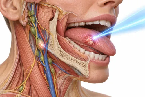
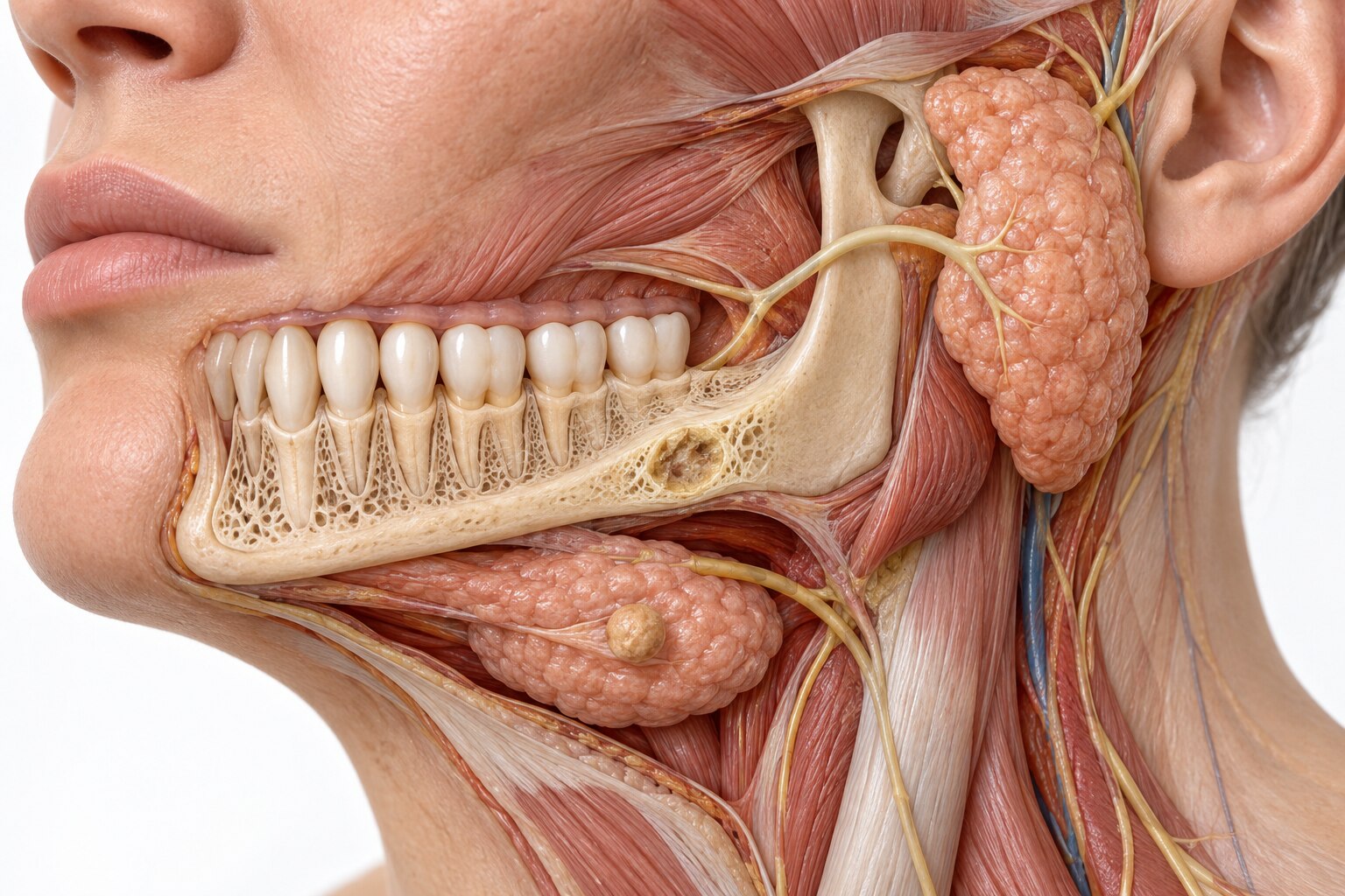
The gland’s location often predicts where you will notice swelling:
- Parotid: in front of and below the ear; a lump appears near the ear or cheek.
- Submandibular: beneath the jaw; disease can swell under the jaw or in the mouth’s floor.
- Sublingual: beneath the tongue; swelling may lift or bulge the area under the tongue.
Pain or enlargement that returns during meals suggests saliva-flow obstruction, such as a stone or narrowed duct. Food stimulates saliva, but the blockage prevents it from draining normally.
A parotid mass needs prompt assessment if it grows quickly, feels fixed, causes persistent pain or numbness, or is accompanied by facial weakness. New reduced facial movement is a high-risk sign, not a routine feature of a simple cyst or benign adenoma.
Salivary gland evaluation in Chennai starts with your history and examination, then selects imaging for the question:
- Ultrasound: an accessible parotid or submandibular mass; it can guide fine-needle aspiration.
- Contrast CT: deep extension, bone involvement or acute infection.
- MRI: deep-lobe parotid disease, skull-base involvement, nerve spread and soft-tissue extent.
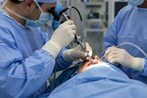
Fine-needle aspiration is usually preferred for an undiagnosed major-gland mass. If it is nondiagnostic, that does not prove the mass is benign; repeat image-guided sampling or core biopsy may be needed. A salivary gland and jaw pathology specialist in Chennai helps when symptoms or imaging cannot be confidently classified.
Why are jaw lesions referred beyond routine dental treatment?
Referral beyond routine dental treatment is needed when the cause, extent or behaviour of a jaw problem cannot be explained confidently by teeth or gums alone. A jaw lesion specialist in Chennai evaluates the finding before extraction, repeated antibiotics or surgery hides the diagnosis.
- An X-ray shows an unclassified lesion, including one found incidentally on a panoramic radiograph.
- A tooth loosens or shifts without periodontal disease explaining it.
- An extraction socket fails to heal, or pain and swelling continue after dental treatment.
- Facial asymmetry, rapid enlargement, unexplained bleeding or restricted mouth opening develops.
- Numbness affects the lower lip, chin or face.
Odontogenic cysts and tumours arise from tooth-forming tissues. Infection can destroy bone, while fibro-osseous disease, metastatic disease and primary bone cancers can create different radiographic patterns. Pain does not separate harmless from serious disease: some benign lesions are painless, while infection and malignancy can both hurt.
A maxilla pathology specialist plans carefully when a lesion approaches the maxillary sinus, nasal cavity, orbit, palate or skull base. The maxilla has less anatomical separation than the mandible, so cross-sectional imaging and coordinated dental, maxillofacial, ENT, oncology or reconstructive planning may be needed before biopsy or surgery.
Treating every radiographic shadow as a dental problem risks sampling the wrong area or compromising later treatment.
Which scans and records help establish the diagnosis?
The right scan answers a specific diagnostic question; no single test covers every salivary gland or jaw problem.
| Test | Main question answered | Best use |
|---|---|---|
| Panoramic radiography | What is the initial overview? | Both jaws, teeth, and an unexpected lesion |
| CBCT | What is the fine bony anatomy? | Teeth, cortical bone, lesion borders, and the mandibular canal |
| Medical CT | Has disease destroyed bone or extended deeply? | Bone destruction, deep extension, spread, or acute infection |
| Ultrasound | Is an accessible gland mass present, and can it be sampled? | Superficial parotid or submandibular masses; image-guided fine-needle aspiration |
| MRI | How far does soft-tissue disease extend? | Deep-lobe parotid disease, skull-base involvement, and perineural spread |
The treating team selects imaging according to the suspected lesion. A normal dental X-ray does not exclude salivary disease, and a normal ultrasound does not reliably exclude a deep-lobe or skull-base process. Persistent symptoms with unrevealing imaging need reassessment, often with CT or MRI.
For salivary gland evaluation in Chennai or mandible lesion treatment in Chennai, bring:
- Panoramic X-rays, CBCT, CT or MRI images and reports
- Biopsy slides or reports and dental records
- Medication and allergy lists
- Details of smoking, chewing tobacco, or other exposure
Ultrasound-guided fine-needle aspiration is commonly the first tissue test for a major salivary-gland mass. If the sample is nondiagnostic, repeat image-guided aspiration or a core biopsy may follow; a nondiagnostic result does not prove benign disease.
How does the diagnosis determine treatment and specialist care?
After referral, treatment is chosen from the diagnosis and expected function, not from an image alone. A scan cannot reliably distinguish a cyst, infection, locally aggressive tumour or cancer without clinical assessment and, when indicated, tissue sampling.
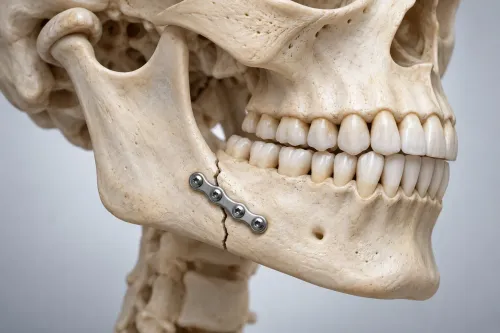
1. Confirm the diagnosis. For a mandible lesion, review its size, location, cortical perforation, teeth involved, relationship to the mandibular nerve and recurrence risk. These findings guide observation, decompression, enucleation, curettage, marginal resection, segmental resection or oncologic treatment.
A maxilla pathology specialist is important when disease approaches the sinus, nasal cavity, orbit, palate or skull base.
2. Match salivary treatment to the cause. Obstruction, inflammation, benign disease and malignancy require different plans. Do not remove an undiagnosed salivary mass automatically; request appropriate imaging, pathology review and tissue assessment first.
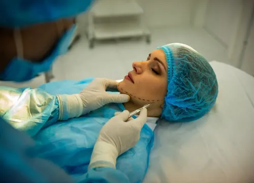
3. Plan cancer care as a team. Suspected cancer may require resection, neck management, radiotherapy, dental rehabilitation and reconstruction. At The Face Clinic, coordinated pathology, maxillofacial and reconstructive decision-making can help when these choices overlap in Chennai.
Before agreeing to surgery, ask:
- Has the pathology been reviewed, and how will margins be assessed?
- Can the facial nerve be preserved, and what would require sacrifice?
- What reconstruction is planned, if any?
- How could treatment affect speech, swallowing, sensation, dental bite and jaw continuity?
A salivary gland and jaw pathology specialist in Chennai should explain alternatives, risks, recurrence, expected function and the reason for each proposed step. That documented explanation is the useful starting point for mandible lesion treatment in Chennai.
Related service
Salivary Gland & Jaw Pathology Management We treat a wide spectrum of benign and malignant lesions involving the salivary glands, maxilla, mandible, and associated soft tissues. View service → |
Frequently asked questions
Which symptoms mean you need specialist assessment?
Seek assessment for a cheek, jaw or neck lump lasting two weeks or longer, unexplained swelling, persistent pain, numbness, difficulty opening your mouth, loose teeth or a non-healing mouth sore.
How can symptoms point to the affected salivary gland?
Swelling or pain that increases during meals suggests a salivary duct blockage. The location also helps: the parotid lies near the ear and cheek, the submandibular gland beneath the jaw, and the sublingual gland under the tongue.
Why are jaw lesions referred beyond routine dental treatment?
A jaw lesion can involve bone, nerves, teeth or nearby soft tissue. Specialist assessment helps distinguish cysts, infections, benign tumours and cancers before treatment is chosen.
Which scans and records help establish the diagnosis?
Bring dental X-rays, panoramic radiographs, CBCT or CT scans, MRI reports, biopsy results, medication details and earlier treatment records. The specialist may request imaging or tissue sampling.
How does diagnosis determine treatment and specialist care?
Treatment depends on the lesion’s type, size, location and behaviour. Options include monitoring, medication, drainage, biopsy, removal or wider surgery, with dental, maxillofacial, reconstructive or cancer specialists involved when needed.
Keywords